how do they x ray babies hips
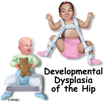
The babys legs have differences in their lengths or appearances. Developmental dysplasia of the hip.
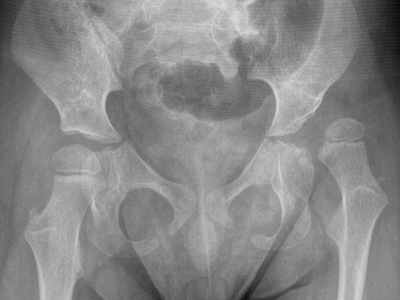
Developmental Dysplasia Of The Hip In Children Eorthopod Com
A hip X-ray is a test that produces an image of the anatomy of your hip.
. When A Young Baby Has A Fever It Can Mean That There Is A Serious. Youll be asked to. Your healthcare provider may use hip X-rays to diagnose and treat health conditions involving your hips.
This is necessary to make the diagnosis or to be sure the hip is normal. This image shows the soft. Your baby will be placed on a table and positioned depending on which body area needs an x-ray.
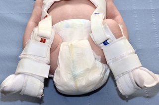
Its a cast that goes around both hips and down the leg to keep the hips. Totaleclipse 07092007 1731. It s sometimes called congenital.
A hip X-ray radiograph is a medical imaging test that creates a. An ultrasound machine sends sound waves into the hip area and images are recorded on a computer. During the examination an X-ray machine sends a beam of radiation through.
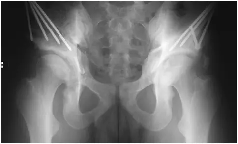
Lying on his back one leg in the knee bends at an angle. If it persists they may put on a spica cast. You will go in the room with him he will need to be stripped from the waist down they will take x-rays of him flat on his back legs dead straight.
Two tests are performed called the barlow and ortolani. The doctor hears or feels a hip click when moving the infants thigh outward during a routine checkup. The main pathology is.
How do they x ray babies uk are a topic that is being searched for and liked by netizens today. If she does have it they may try to brace it first. For babies 4 months of age or older and children x-rays are performed when hip dysplasia is suspected.
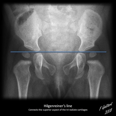
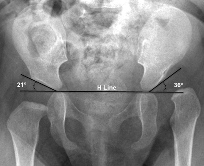
Value How Do They X Ray Babies Hips 2022. If an X-ray of the hip joints is performed according to Launstein Lauenstein then the patients position looks like this. Two tests are performed called the.
A hip X-ray is a safe and painless test that uses a small amount of radiation to make images of the hip joints where the legs attach to the pelvis. Its sometimes called congenital dislocation of the hip or hip dysplasia. An X-ray of the pelvis focuses specifically on the area between your hips that holds many of your reproductive and digestive organs.
During the examination an X-ray machine sends a beam of radiation through the pelvic bones and hip joints and an image is recorded on a computer or special film. Your How do they x ray babies uk images are available.
Developmental Dysplasia Of The Hip Radiology Reference Article Radiopaedia Org

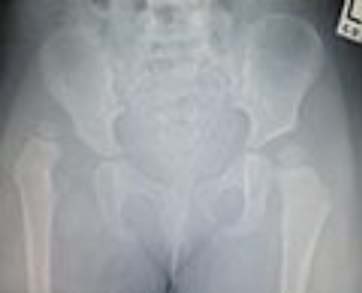
Pelvic X Ray Different Ages Radiology Case Radiopaedia Org

Infant Diagnosis International Hip Dysplasia Institute

Diagnosis Of Hip Dysplasia Healthy Hips Australiahealthy Hips Australia
Developmental Dysplasia Of The Hip Nhs

What Are The Signs Of Hip Dysplasia In Babies And Adults Treatment

60 Xray Of Child Hip Stock Photos Pictures Royalty Free Images Istock
Hip Dysplasia Information Symptoms Diagnosis Treatment
Hip Surveillance Helps Identify Dislocations In Children With Cerebral Palsy Children S National
Imaging Of Developmental Dysplasia Of The Hip Ultrasound Radiography And Magnetic Resonance Imaging Springerlink

Hip Subluxation Dislocation And Surveillance In Children With Cerebral Palsy Cp

Hip I Developmental Dysplasia Of The Hip Musculoskeletal Key

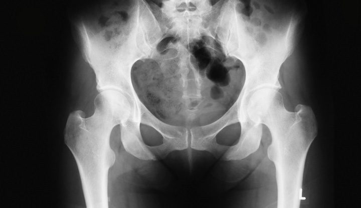
X Ray Of Hip Dysplasia Wikipedia
Total Hip Arthroplasty For Dysplasia And Congenital Disease Of The Hip Review Paper
Pelvis X Ray Purpose Procedure Risks

Hip Dysplasia Should My Child Be Screened Uva Radiology

How To Prevent Hip Dysplasia In Babies Physical Therapy Center
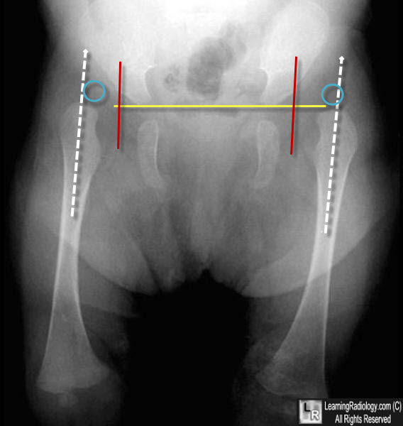
Learningradiology Developmental Dislocation Dysplasia Of The Hip

Developmental Dysplasia Of The Hip Johns Hopkins Medicine